Leave message
Can’t find what you’re looking for?
Fill out this form to inquire about our custom protein services!
Inquire about our Custom Services >>


> MHC Complex Custom Service 
![]() Eukaryotic expression platform for a eukaryotic-based conformation
Eukaryotic expression platform for a eukaryotic-based conformation
![]() Professional MHC-peptide complex R&D technology platform
Professional MHC-peptide complex R&D technology platform
![]() Various types of MHC I and antigens are available
Various types of MHC I and antigens are available
![]() Mature bioactivity verification platform: SPR/ELISA/FACS and more
Mature bioactivity verification platform: SPR/ELISA/FACS and more
![]() Co-expression of heterodimer results in a more natural conformation
Co-expression of heterodimer results in a more natural conformation
![]() Monomer/Tetramer forms
Monomer/Tetramer forms
![]() FITC/PE/APC/Biotin-labeled are optional
FITC/PE/APC/Biotin-labeled are optional


| MHC Allele | |||||||
|---|---|---|---|---|---|---|---|
| Human classical MHC class I | Human classical MHC II | Mouse | Monkey | ||||
| Antigens |
|---|
Bioactivity
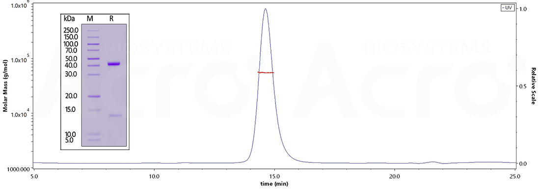
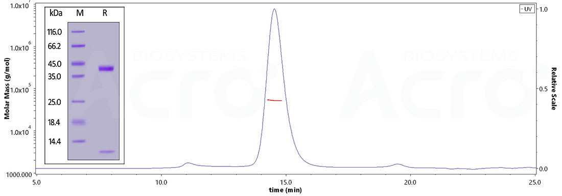
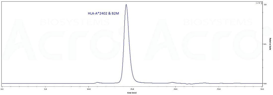
Homogeneity
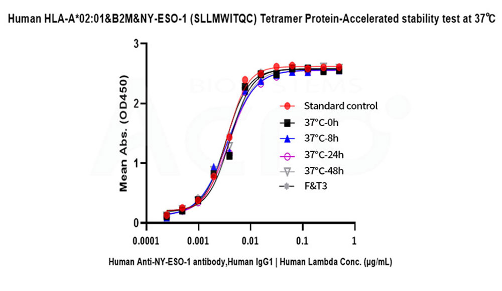
Stability
Competitive Data
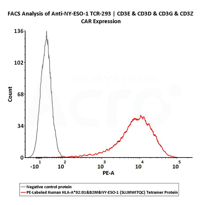
PE-Labeled Human HLA-A*02:01&B2M&NY-ESO-1 (SLLMWITQC) Tetramer Protein (Cat. No. HL1-HP2E5) and negative control protein were first diluted in a 1:25 dilution ratio with FACS buffer. Staining was performed using 100 µL of working solution to 5e5 anti-NY-ESO-1 TCR-293 cells. Binding activity was evaluated based on PE signal intensity (QC tested).
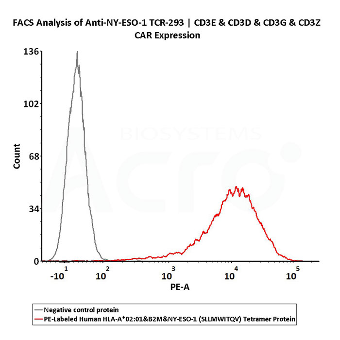
PE-Labeled Human HLA-A*02:01&B2M&NY-ESO-1 (SLLMWITQV) Tetramer Protein (Cat. No. HL1-HP2E6) and negative control protein were first diluted in a 1:25 dilution ratio with FACS buffer. Staining was performed using 100 µL of working solution to 5e5 anti-NY-ESO-1 TCR-293 cells. Binding activity was evaluated based on PE signal intensity (QC tested).
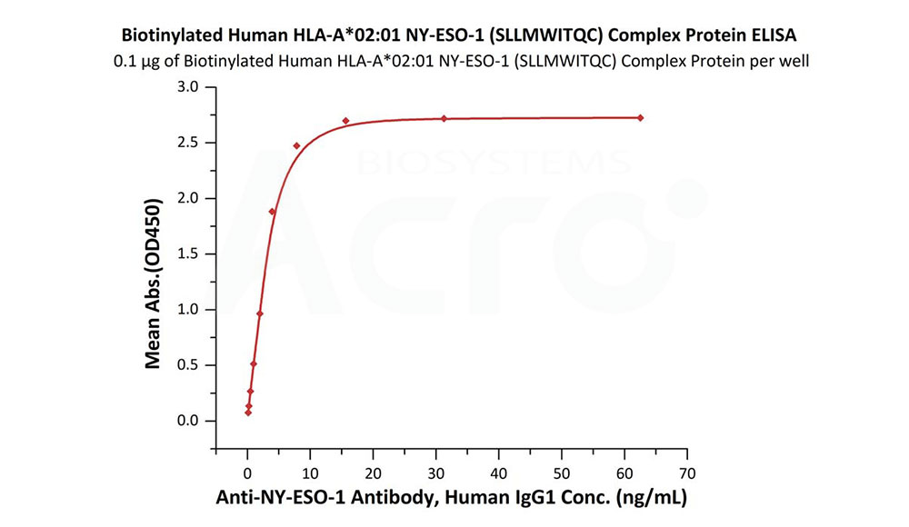
Immobilized Biotinylated Human HLA-A*02:01&B2M&NY-ESO-1 (SLLMWITQC) Complex Protein (Cat. No. HL1-H82E6) at 1 μg/mL (100 μL/well) on streptavidin (Cat. No. STN-N5116) precoated (0.5 μg/well) plate can bind Anti-NY-ESO-1 Antibody, Human IgG1 with a linear range of 0.1-8 ng/mL (QC tested).
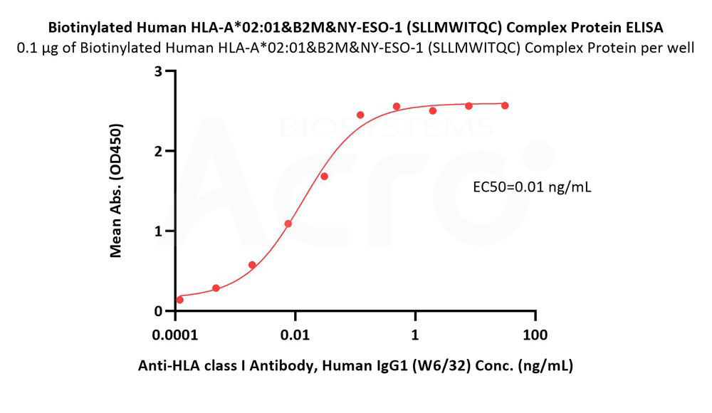
Immobilized Biotinylated Human HLA-A*02:01&B2M&NY-ESO-1 (SLLMWITQC) Complex Protein (Cat. No. HL1-H82E6) at 1 μg/mL (100 μL/well) on streptavidin (Cat. No. STN-N5116) precoated (0.5 μg/well) plate can bind Anti-HLA class I Antibody, Human IgG1 (W6/32) with a linear range of 0.1-1 ng/mL (Routinely tested).
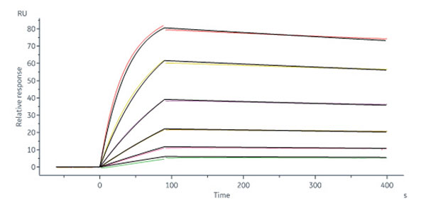
Anti-HLA class I Antibody, Human IgG1 (W6/32) captured on Protein A Chip can bind Human HLA-A*11:01&B2M&KRASG12D (VVGADGVGK) Complex Protein (Cat. No. HLD-H52H4) with an affinity constant of 1.06 nM as determined in a SPR assay (Biacore 8K) (Routinely tested).
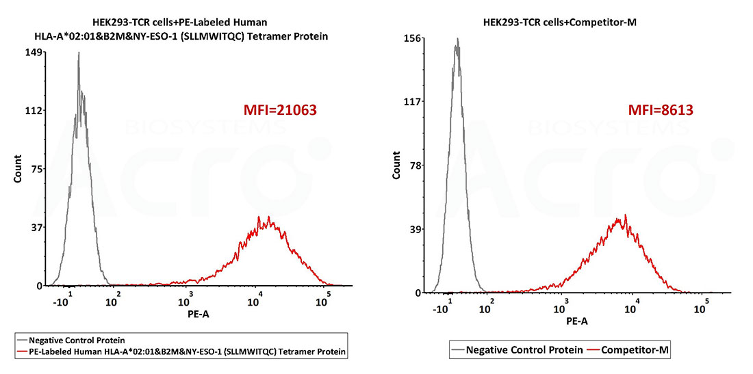
Performance of PE-labeled Human HLA-A*02:01&B2M&NY-ESO-1 (SLLMWITQC) Tetramer from ACROBiosystems (Cat. No. HL1-HP2E5) and other brand were evaluated by flow cytometry under the manufacturer’s recommended conditions. ACROBiosystems’ PE-Labeled NY-ESO-1 Tetramer Protein shows 3x higher MFI signal strength than competitor M, revealing much better TCR binding.
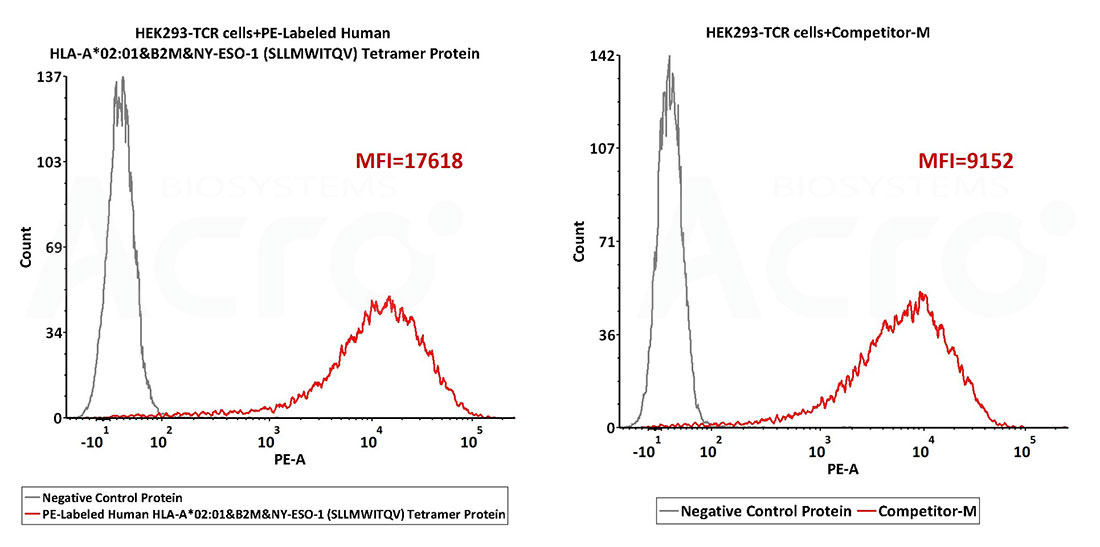
Performance of PE-labeled Human HLA-A*02:01&B2M&NY-ESO-1 (SLLMWITQV) Tetramer from ACROBiosystems (Cat. No. HL1-HP2E6) and other brand were evaluated by flow cytometry under the manufacturer’s recommended conditions. ACROBiosystems’ PE-Labeled NY-ESO-1 Tetramer Protein shows 2x higher MFI signal strength than competitor M, revealing much better TCR binding.


This web search service is supported by Google Inc.


 A-Z
A-Z